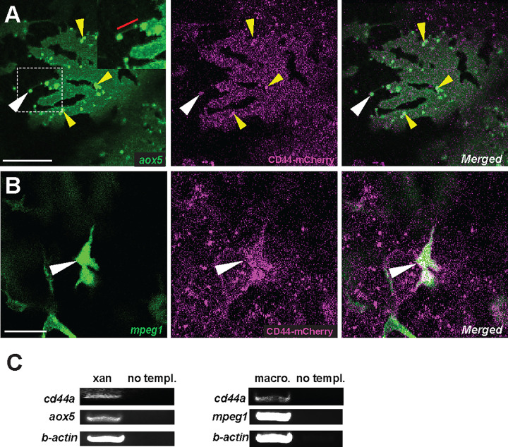
Figure 2. Localization of CD44a protein in xanthoblasts and macrophages.
(A) CD44-mCherry expression across the xanthoblasts cell membrane. CD44 is also notably present in the airineme vesicle (white arrowhead) and in the airineme blebs (yellow arrowheads). Airineme filament is indicated with a red bar in the inset. (B) CD44-mCherry expression in macrophages (white arrowheads). (C) RT-PCR for cd44a in isolated xanthophores (xan) and macrophages, and no template control. Scale bars represent 20μm (A, B).

