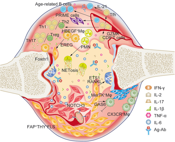
FIGURE 2.

The immune microenvironment composed of different pathogenic or protective cell subsets in RA. The imbalance of Th1/Th2 and Th17/Treg is involved in RA. GZMK+CD8+ T cells produce large amounts of IFN‐γ; CD4+PD1+CXCR5− Tfh cells secrete IL‐21 to activated B lymphocytes; PRIME cells derived from B lymphocytes can predict the flare of RA; aging‐associated B cells have been found to be pathogenic in RA; macrophages provide many regulators such as Foxm1 and ETS1 to activate osteoclasts, and EREG to stimulate FLS as well; FAP+THY+FLS exhibits autoimmune phenotype, and after receiving NOTCH3 signal from endothelial cells, they became invasive. However, some subsets of macrophages are protective, such as MerTK+ Mφ and CX3CR+ Mφ.
