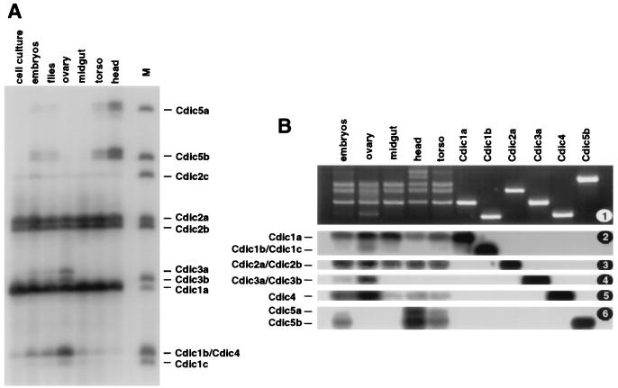
FIG. 7.
Tissue specificity of Cdic isoforms. RT-PCR fragments were amplified across the variable region of Cdic transcripts. (A) PCR fragments were labeled at one DNA strand with 32P and separated in a 5% acrylamide sequencing gel. The source of the RNA is indicated at the top. Lane M contains marker fragments generated from the of cDNA clones representing Cdic isoforms. (B) PCR fragments were separated in a 3% agarose gel (1) and, after Southern transfer, hybridized with oligonucleotides "iso2" (2), "iso2‴ (3), "iso3" (4), "iso4" (5), and "iso5" (6). The source of the RNA is indicated at the top; individual cDNA clones Cdic1a, Cdic1b, Cdic2a, Cdic3a, Cdic4, and Cdic5b were used to generate the marker fragments in the six right-hand lanes.

