Figure 5.
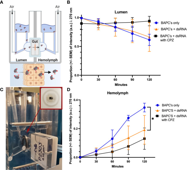
Cellular uptake study. Mechanism of Rh–BAPCs and Rh–BAPC-dsRNA cellular uptake by P. japonica midgut cells. (A, B) graphical representation and actual set up of Ussing chamber used for ex vivo analysis of BAPC-dsRNA complexes uptake and transport across P. japonica midgut tissue. (C) Mean relative fluorescence of Rh-BAPCs complexes on luminal side buffer and (D) Mean relative fluorescence of Rh-BAPCs complexes on hemolymph side buffer over 2 hr. Differences between values were compared by two-way ANOVA using Tukey as post-test. Statistical significance: (*) P< 0.05.

