
Fig. 3. GRK recruitment to CXCR3 in G protein–deficient cells.
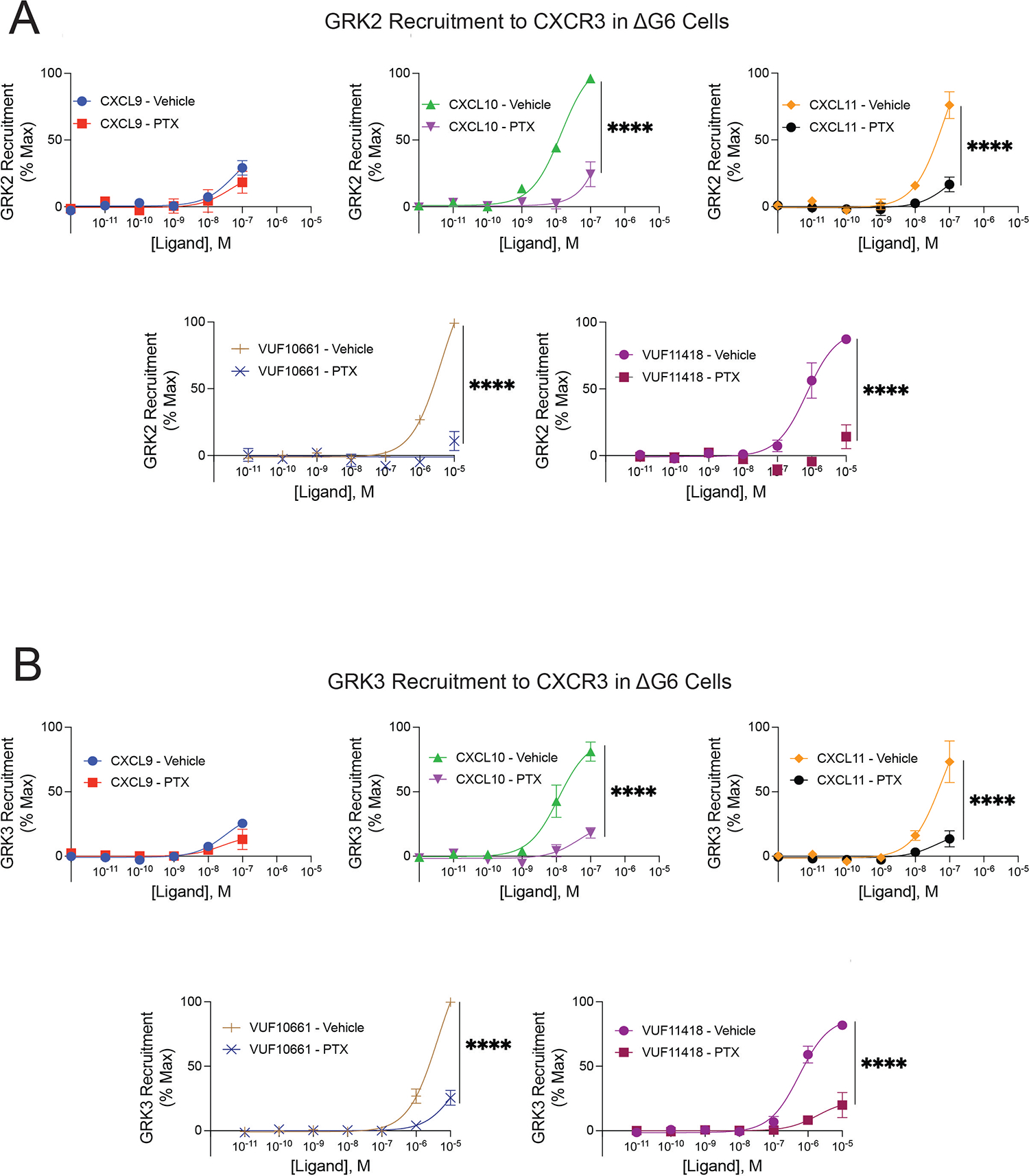
Agonist concentration-response of (A) GRK2 and (B) GRK3 recruitment to CXCR3 with and without pretreatment with 200ng/mL pertussis toxin (PTX) in ΔG6 knockout cells treated with CXCL9, CXCL10, CXCL11, VUF10661 or VUF11418. GRK recruitment was measured using a split luciferase assay involving LgBiT-tagged GRK2 or GRK3 and CXCR3-SmBiT. Data shown are the mean ± SEM of n=3 independent experiments, significance testing by one-way ANOVA with Šidák post hoc testing comparing luminescent signal at maximum dose between treatment conditions (vehicle vs. PTX). Concentration-response curves are normalized to maximum signal observed across all ligands. *P<0.05, **P<0.005, ***P<0.0005, ****P<0.0001.