FIG. 6.
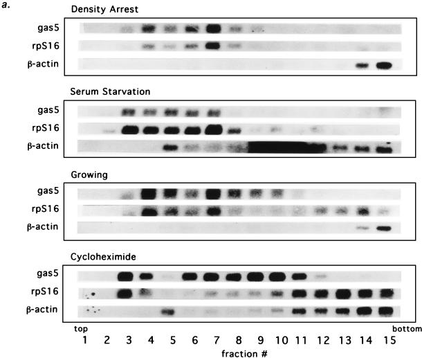
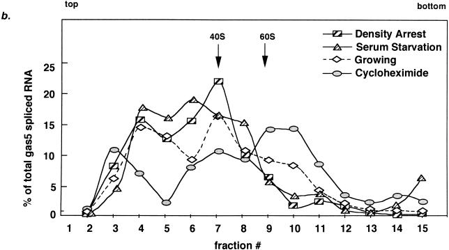
Spliced gas5 RNA shifts away from ribosomes during growth arrest. (a) Northern blots of RNA from 10 to 50% sucrose gradient fractions of extracts from NIH 3T3 cells density arrested, serum starved, growing, or treated with cycloheximide. The distributions of spliced gas5 RNA and rpS16 and β-actin mRNAs, along with 18S and 28S rRNAs (data not shown), were analyzed. The top, bottom, and fraction numbers for each gradient are indicated. The dark smear in fractions 9 to 12 of the β-actin profile during serum starvation is an artifact introduced during the blotting procedure; note that this profile otherwise mimics the β-actin profile during cycloheximide treatment. (b) Quantitation of Northern signals (by PhosphorImaging) of spliced gas5 RNA from density-arrested cells, serum-starved cells, growing cells, and cells treated with cycloheximide. The positions of ribosomal small subunits (40S) and large subunits (60S) were determined from the profiles of 18S and 28S rRNAs. For each fraction, the percentage of gas5 was determined with respect to the total amount of gas5 RNA in the gradient. The dotted line indicates that the normalized level of gas5 RNA in growing cells cannot be compared directly to that in density-arrested and cycloheximide-treated cells, since a portion of the spliced gas5 RNA (presumably that associated with ribosomes) has been degraded.


