Fig. 2.

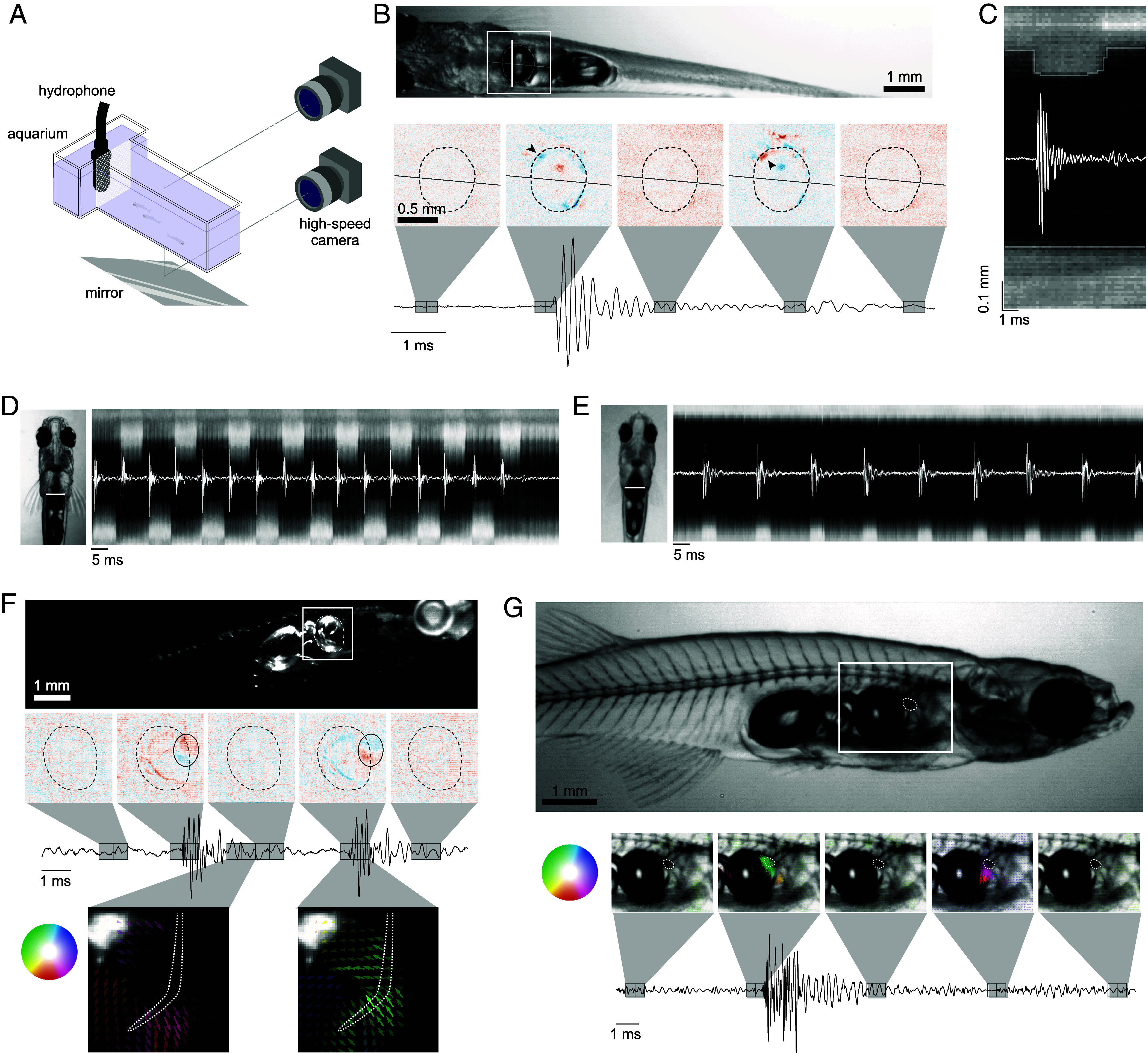
High-speed video reveals ultrafast motion during sound production. (A) High-speed video setup. 3 to 4 fish, including at least one male were placed inside the tank, separated from the hydrophone by a piece of mesh. The camera was moved to image the tank either from the side or from below with the use of a mirror. (B) A video of a male (Movie S2) (example frame above) was registered and the difference between consecutive frames displays the unilateral compression and relaxation of the anterior part of the swim bladder (black arrow heads) which corresponded with the onset of the pulse and after-pulse, respectively. (C) The pixels along the vertical white line in B were plotted over one pulse to show the dynamics of the swim bladder during sound production. The compression occurred over less than 0.125 ms (faster than one frame) whereas the relaxation occurred over about 0.5 ms. (D) A high-speed video from below (Movie S5) was registered and the pixels along the vertical white line in the example frame were plotted over a burst with a pulse rate of 120 Hz. The compression of the swim bladder alternated between the left and right side of the fish. (E) The same as in (D) except for a 60 Hz burst (Movie S6). The swim bladder compression is unilateral in this case. (F) A video of a male viewed from the side (Movie S7) (example frame above) with the illumination from above was registered and the difference between consecutive frames revealed that only a small area of the anterior part of the swim bladder was compressed, outlined with a black oval. In this view, we also observed rostral movement of the rib before the pulse (Bottom Left), and caudal movement after (Bottom Right). (G) A video of a male from the side (Movie S8) (example frame above), with backlight illumination. The optical flow between consecutive frames shows the motion of the cartilage, outlined with a white dotted line. The color and opacity of the arrows corresponds to the direction and amplitude of the motion respectively.
