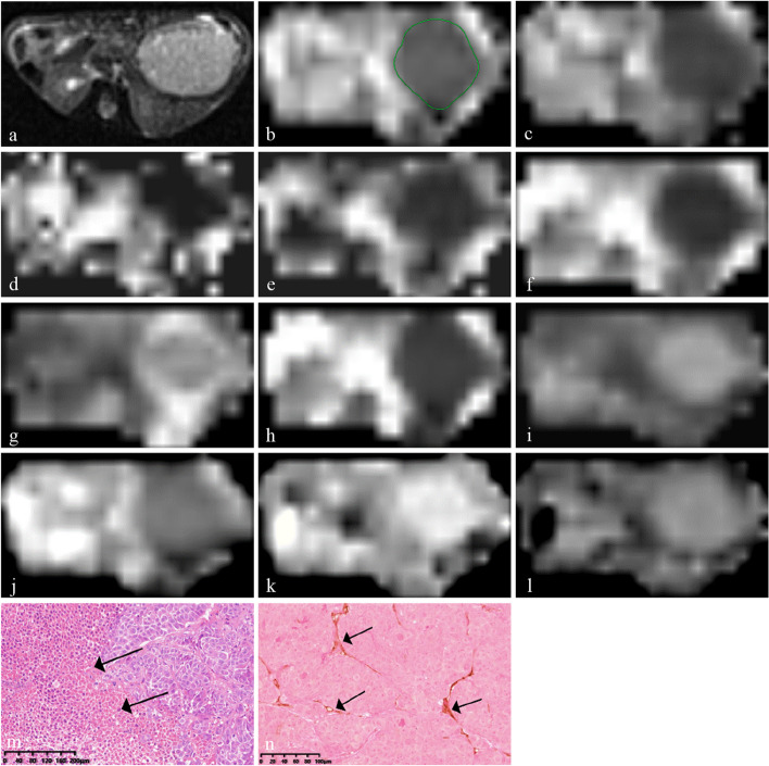
Fig. 1.
Multi-b-value DWI images and corresponding histopathological images of hepatocellular carcinoma in the bufalin plus sorafenib treatment group. a A transverse T2-weighted image shows the slice with the maximum tumor diameter. b Apparent diffusion coefficient map for outlining the tumor. c–i Dt, Dp, f, mean diffusivity, mean kurtosis, distributed diffusion coefficient, and α tumor maps and (j–l) D, β, and µ tumor maps. m Hematoxylin-eosin staining showing patchy necrosis (black arrows, ×10). n Anti-CD31 staining image showing sparse microvessels (black arrows, ×20)

