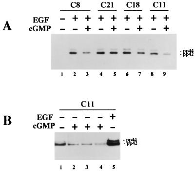
FIG. 4.
Assessment of MAP kinase phosphorylation by using a phospho-MAP kinase-specific antibody. Cells were serum starved in the absence of zinc and treated with 8-pCPT-cGMP and/or EGF as indicated. Western blots were prepared as described for Fig. 3 but were developed with an antibody specific for the dually phosphorylated, active form of MAP kinase as described in Materials and Methods. (A) EGF (10 ng/ml; lanes 2 to 9) was added for the last 5 min prior to harvesting, and 8-pCPT-cGMP (500 μM; lanes 3, 5, 7, and 9) was added 30 min prior to addition of EGF. Clone C8, C18, and C11 cells express significant amounts of G-kinase activity, whereas C21 cells are G-kinase deficient. (B) Clone C11 cells were left untreated (lane 1) or were treated with 500 μM 8-pCPT-cGMP for 15 (lane 2), 30 (lane 3), or 60 (lane 4) min prior to harvesting. Lane 5, cells treated with EGF (10 ng/ml) for 5 min. Exposure times were 15 s (A) and 5 min (B).

