
Fig. 2.
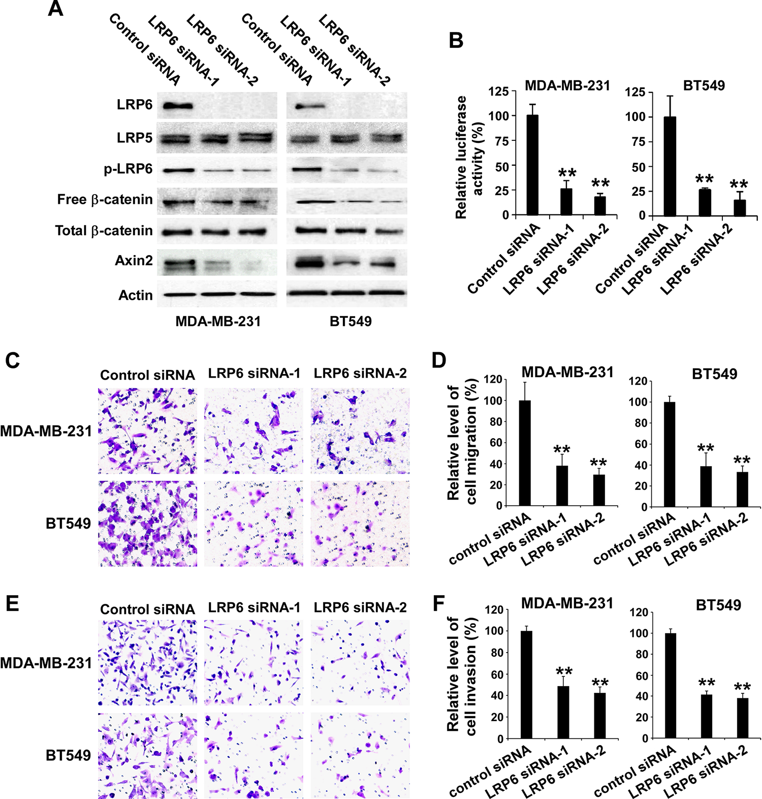
Depletion of LRP6 results in inhibition of Wnt/β-catenin signaling in TNBC cells and suppression of TNBC cell migration and invasion. (A) MDA-MB-231 and BT549 cells in 6-well plates were transiently transfected with 50 nM of LRP6 siRNA-1, LRP6 siRNA-2 or control siRNA. After incubation of 48 h, the levels of human LRP6, LRP5, phospho-LRP6 (p-LRP6), cytosolic free human β-catenin, total cellular human β-catenin, and axin2 were examined by Western blotting. All the samples were also probed with anti-human actin antibody to verify equal loading. (B) MDA-MB-231 and BT549 cells in 24-well plates were transiently transfected with 50 nM of LRP6 siRNA-1, LRP6 siRNA-2 or control siRNA. After incubation of 24 h, cells were transfected with the Super8XTOPFlash luciferase construct and β-galactosidaseexpressing vector in each well. The luciferase activity was then measured 24 h later with normalization to the activity of the β-galactosidase. Values are the average of triple determinations with the s.d. indicated by error bars. (C-F) MDA-MB-231 and BT549 cells were transiently transfected with 50 nM of LRP6 siRNA-1, LRP6 siRNA-2 or control siRNA. After incubation of 48 h, the transwell migration assay (C, D) and Matrigel invasion assay (E, F) were performed. (C) Representative images of cell migration. (D) Relative levels of cell migration are significantly decreased following LRP6 knockdown in MDA-MB-231 and BT549 cells. (E) Representative images of cell invasion. (F) Relative levels of cell invasion are significantly decreased following LRP6 knockdown in MDA-MB-231 and BT549 cells. Data shown are representative of three independent experiments. All the values are the average of quadruplicate determinations with the s.d. indicated by error bars. **P < 0.01 versus control cells.