
Fig. 5.
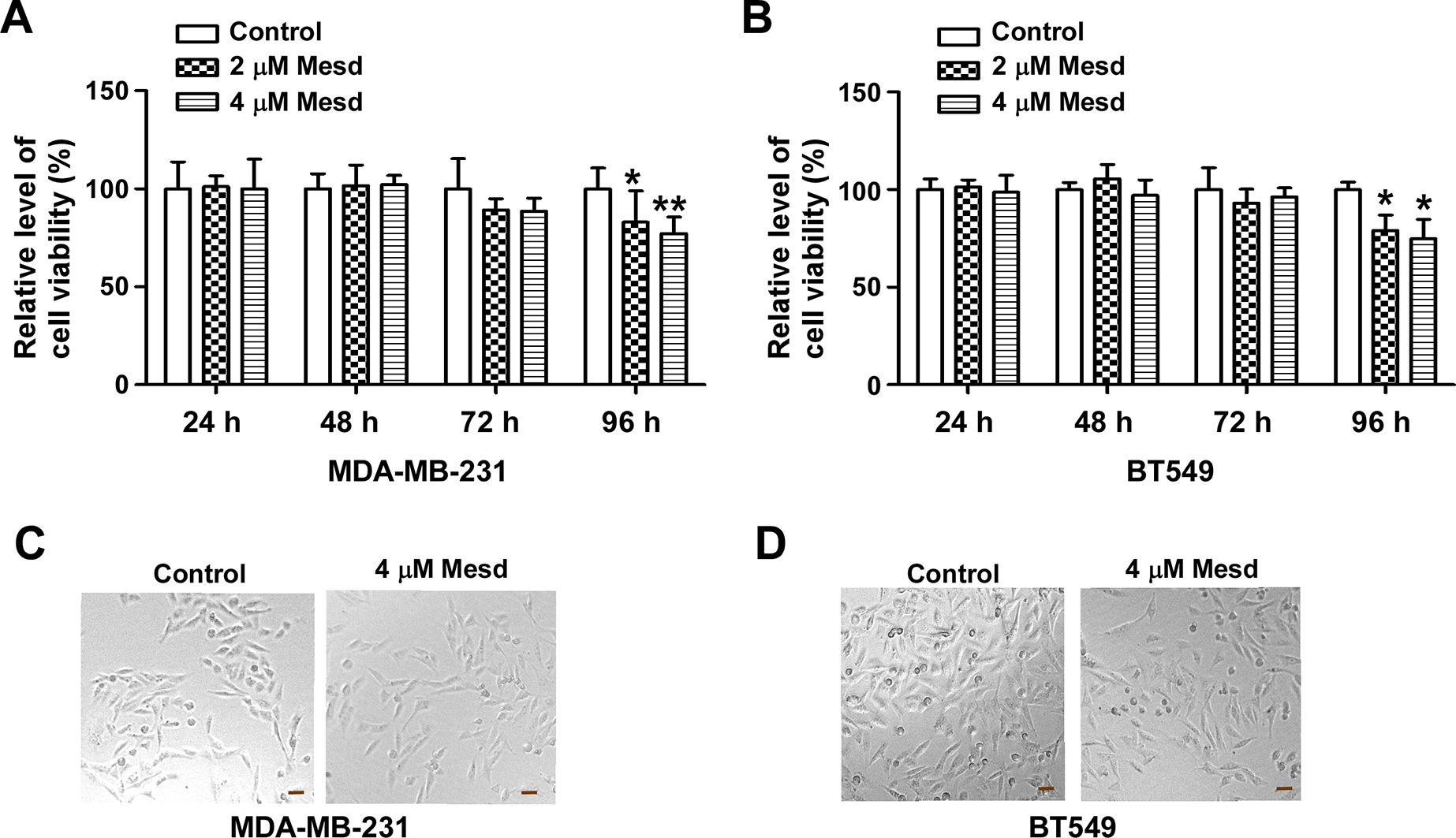
Mesd inhibits TNBC cell proliferation/viability. (A, B) MDA-MB-231 and BT549 cells in 96-well plates were treated with Mesd at the indicated concentrations. After incubation for 24, 48, 72 or 96 h, cell viability was measured by the Cell Titer Glo Assay system. Data shown are representative of three independent experiments. All the values are the average of triplicate determinations with the s.d. indicated by error bars. *P < 0.05, **P < 0.01 versus control cells. (C, D) MDA-MB-231 and BT549 cells were treated with Mesd (4 μM) for 96 h, and images were taken using phase contrast microscopy. Scale bar: 40 μm.