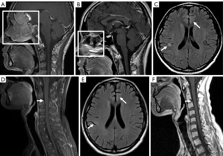
Figure 2.
Changes in magnetic resonance imaging findings of olfactory neuroblastoma. A contrast-enhanced sagittal magnetic resonance T1-weighted sequence of the brain (A) showing an expansile lesion involving the ethmoid sinus, sphenoid sinus, nasal cavity, and frontal lobe with bone destruction. After 6 courses of chemotherapy and immunotherapy, a contrast-enhanced sagittal T1-weighted sequence (B) indicated that the above lesion had obviously diminished. At the same examination, an axial T2-weighted FLAIR sequence (C) revealed some new expansile lesions presenting as a hyperintensity nodule in the right parietal sulcus and a patch in the left frontal lobe. Linear, meningeal-enhanced thickening was observed on T1-weighted, fat-suppression, gadolinium-enhanced spinal magnetic resonance imaging representing leptomeningeal disease (D). The lesion was suspected to be immune encephalitis. After adsorption treatment, the new lesions had almost disappeared in the follow-up axial T2-weighted FLAIR (E) and sagittal T1-weighted contrast-enhanced images (F). Boxes show tumor location, and arrows show intracranial and spinal cord lesions. FLAIR, fluid attenuated inversion recovery.

