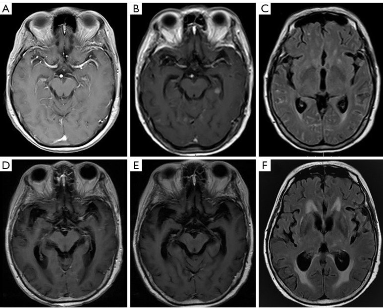
Figure 5.
Positive magnetic resonance imaging findings of meningeal metastasis of lung adenocarcinoma. A 67-year-old female was diagnosed with lung adenocarcinoma. At first, she had a negative imaging finding on a T1-weighted, gadolinium-enhanced sequence (A). After 3 months, contrast-enhanced T1-weighted magnetic resonance imaging showed abnormal meningeal enhancement and nodular enhancement in the left hippocampus (B). More meningeal lesions were found in axial T2-weighted FLAIR (fluid-attenuated inversion recovery), and the gadolinium-enhanced sequence presented multiple linear hyperintensities (C). After radiotherapy, the T1-weighted, gadolinium-enhanced sequence showed that the above meningeal lesions had gradually disappeared (D,E). Conversely, based on the axial T2-weighted FLAIR sequence, hydrocephalus was worse than before (F). FLAIR, fluid attenuated inversion recovery.

