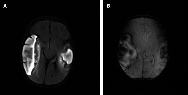
Figure 4.
(A) Axial Ffluid A-attenutaed Iinversion Rrecovery (FLAIR) shows bilateral multiple extensively hyperintense lesions at frontal temporal regions shown on MRI. (B:) Axial Ffast Ffield Eecho (FFE) shows bilateral frontal temporal lesion with blooming artefacts in keeping with areas of cerebral haemorrhages in MRI.

