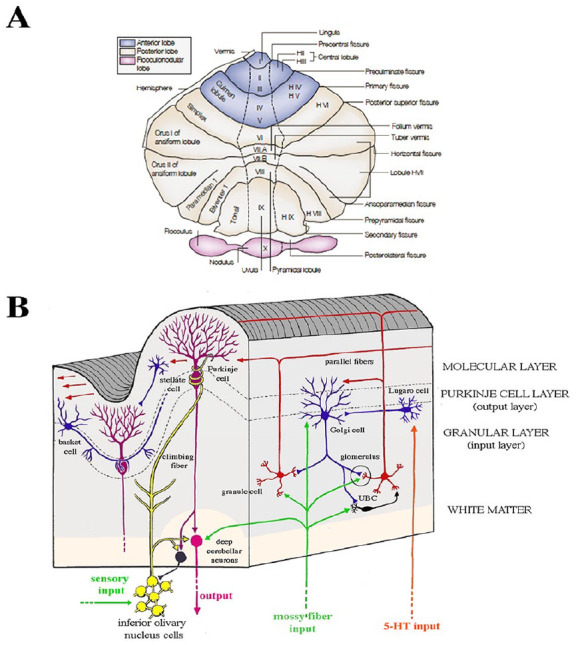
Figure 3.

Macro- and microanatomy of the human cerebellum. (A) Unfolded view of the cerebellar cortex shows lobes, lobules by name and number, and main fissures. Hemispherical lobules are designed by the prefix “H” according to Larsell’s classification and are followed by Roman numerals indicating the corresponding vermian lobules. Adapted from Manni and Petrosini (2004). (B) Cellular and fiber elements of the cerebellar cortex. A vertical section of a single cerebellar folium, in longitudinal and transverse planes, illustrates the general organization of the cerebellar cortex. The cellular architecture of the cerebellar cortex is uniform throughout the folia. Purkinje cells, the sole output of the cerebellar cortex, mainly project to the deep cerebellar nuclei and receive excitatory input on their extensive arborization from a beam of parallel fibers arising from several granule cells and from a single climbing fiber arising from the inferior olive.
