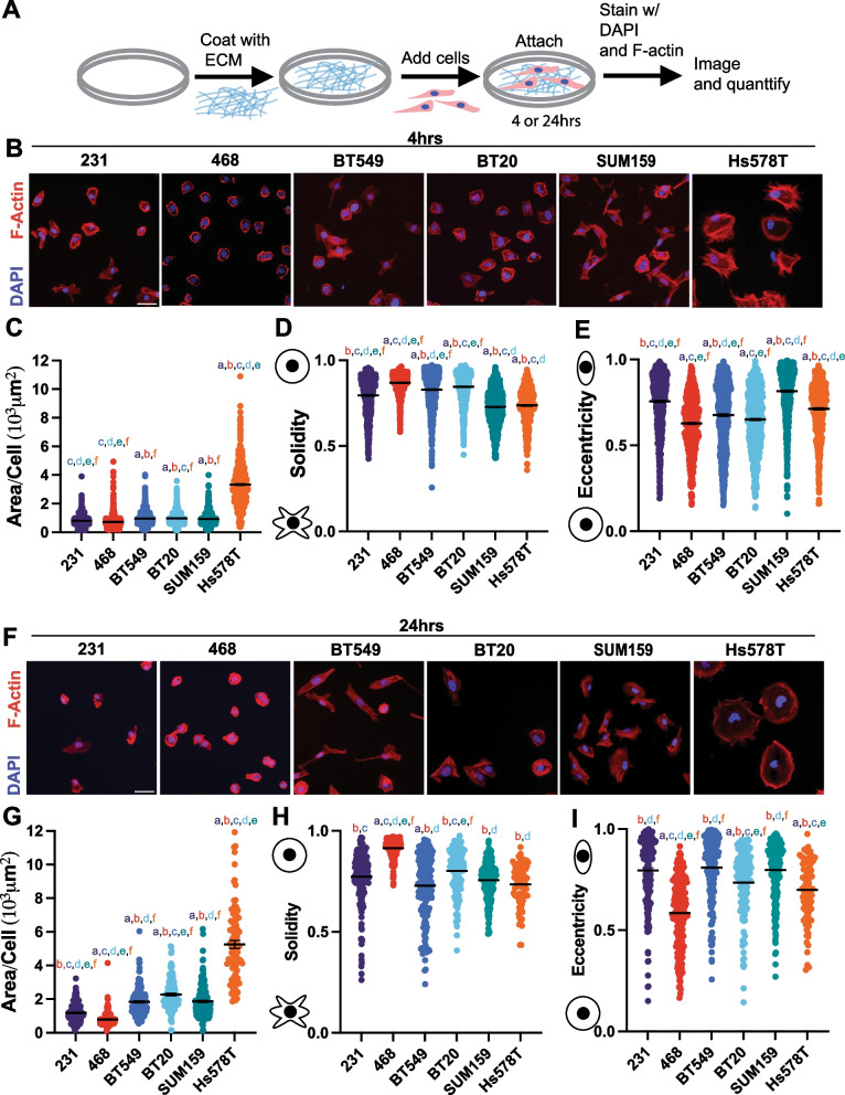
Fig. 4.
Characterization of TNBC cell line morphology. A Schematic depicting experimental procedure. B Representative images of 231, 468, BT549, BT20 SUM159 and Hs578T cell lines plated on Collagen I ECM for 4 h, fixed and stained for nuclei (Blue) and F-actin (red). Scale bar = 50 μm. Quantification of cell shape parameters for each cell line 231 (n = 679 cells), BT549 (n = 799 cells), Hs578T (n = 538 cells), 468 (n = 818 cells), BT20 (n = 882 cells) and SUM159 (n = 892 cells) C Area/Cell, D Solidity, and E Eccentricity. F Representative images of 231, BT549, Hs578T, 468, BT20, and SUM159 cell lines plated on Collagen I for 24 h, fixed and stained for nuclei (Blue) and F-actin (red). Scale bar = 50 μm. Quantification of cell shape parameters for each cell line 231 (n = 178 cells), BT549 (n = 184 cells), Hs578T (n = 91 cells), 468 (n = 205 cells), BT20 (n = 135 cells) and SUM159 (n = 192 cells) G Area/Cell, H Solidity, and I Eccentricity. Data show mean ± SEM. Significance was determined using a one-way ANOVA with Tukey’s multiple comparison test comparing each cell line to every other cell line. Significance (p < 0.05) is denoted by a letter corresponding to each cell line tested, 231 (a), 468 (b), BT549 (c), BT20 (d), SUM159 (e), Hs578T (f)

