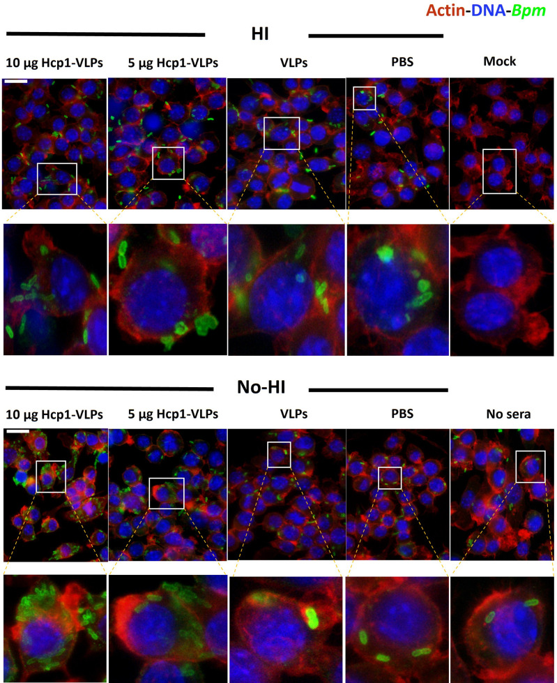
Fig 6.
Fluorescence microscopic analysis of RAW 264.7 macrophage cells after 2 h of incubation with B. pseudomallei K96243 in the presence of HI and no-HI immune sera from PBS, VLPs, 5 or 10 µg Hcp1-VLPs group. Cells incubated with cDMEM with (no sera) or without bacteria (mock) were used as controls. After infection, the cells were fixed with paraformaldehyde, permeabilized with Triton X-100 before incubation with serum from mice vaccinated with B. pseudomallei PBK001. The bacterial cells, macrophage cell nuclei, and actin were examined by a rabbit anti-mouse Alexa Fluor-488, DAPI, and rhodamine phalloidin, respectively. Images were taken from an Olympus BX51 upright fluorescence microscope (60×) then processed using ImageJ software. The scale bar represents 20 µm.

