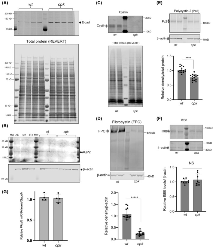
FIGURE 2.

Reduced FPC levels in cpk, compared to wt CCD cells. (A,B) Characterization of wt and cpk CCD cell lines (n = 4). (A) E‐cadherin (E‐cad) expression was tested as epithelial marker. Cell lysates were randomly selected (wt, cpk) from experiments performed in a 6‐month period (n = 4). (B) Aquaporin‐2 (AQP2) expression (CCD marker, top) in randomly selected from wt and cpk cell lysates. AE: airway epithelial cells (positive control), MK: mixed mouse kidney epithelial cells, 3 T3: mouse fibroblast cell line (negative control). β‐actin as loading control (bottom). (C) Cpk cells lack cystin. Cystin was only present in wt cells. Cystin was labeled with rabbit polyclonal anti‐cystin antibody (top). Total protein stain (REVERT) is presented in the lower panel to demonstrate equal loading. (D) Reduced FPC levels in cpk cells compared to wt. 30 μg of total cellular proteins was analyzed by WB using an anti‐FPC C‐terminal, rat monoclonal antibody (representative gel on top). Relative FPC abundance was quantitatively assessed by densitometry and expressed relative to β‐actin (bottom). Experiments were performed using cell culture lysates from different passage numbers (N = 7, n = 14, unpaired t‐test). (E) Pc2 reduction in cpk cells. Pc2 WB using a rabbit polyclonal Pc2 antiserum (top). Pc2 abundance was quantitatively assessed by densitometry and expressed relative to β‐actin. Same cell lysates as for FPC were tested for Pc2 (N = 7, n = 14, ****p < .0001, unpaired t‐test). (F) Ift88 levels are similar in wt and cpk cells. Ift88 was labeled with anti‐Ift88 rabbit polyclonal antiserum. No significant differences were observed based on quantitative assessment of densitometry expressed relative to β‐actin (N = 4, n = 8, NS, unpaired t‐test). Same lysates were tested as for FPC and Pc2. (G) No reduction in relative Pkhd1 mRNA levels in cpk cells compared to wt. Plotted values indicate mRNA levels in four independently derived samples relative to Gapdh1 mRNA (from cell cultures at different passage number, n = 4, NS, unpaired t‐test).
