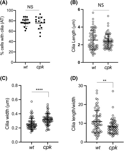
FIGURE 4.

Loss of cystin in cpk cells does not affect cilia formation but leads to altered ciliary architecture. Acetylated tubulin (AT) staining was used to determine cilia number, thickness, and length. (A) Similar percentage of wt and cpk cells develop primary cilia. Cilia development was induced by serum starvation. Each data point represents the percentage of cells with cilia in a field with 22–44 cells in two experiments (wt: n = 492, cpk: n = 533). (B) Cilia length; (C) width (thickness, at the base of the cilia) was measured using ImageJ (see Materials and Methods). Each data point represents measurements of one cilium (n = 61, ****p < .0001). (D) Cilia length/width ratios are plotted from data points presented in (B) and (C) (n = 61, **: p = .0027).
