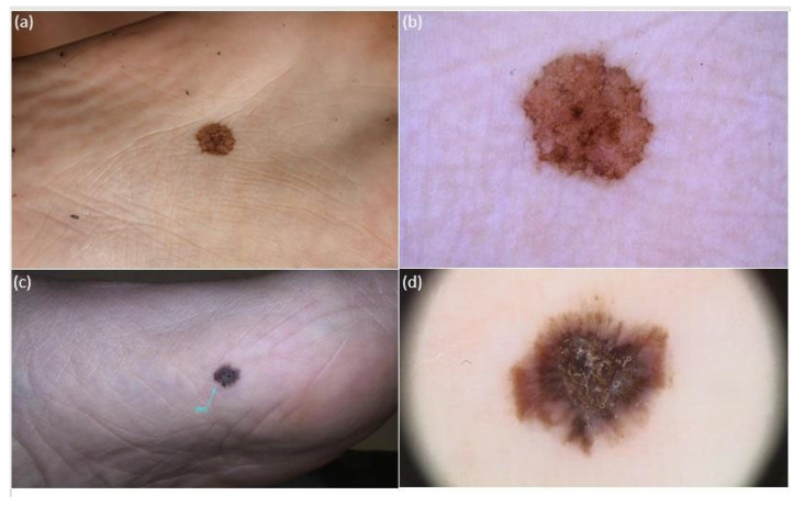
Figure 2.
Clinical and dermoscopic (polarized light, 20×) appearance of 2 atypical melanocytic plantar lesions (aMPPLs) of the sole, localized at the central eminence (a,b) and anterior–medial eminence (c,d). Both lesions appear as brownish roundish pigmented macules with clear-cut borders and non-homogenous pigmentation, similar diameter and multiple colors, and irregular blotches observed under dermoscopy; however, the lesion of the central eminence was an atypical nevus of 12 mm in a 20-year-old female (a,b), while the lesion on the anterior–medial eminence was an early melanoma (pt1a) of 13.6 mm in a 63-year-old male (c), with additional dermoscopic features of a hyperkeratosic component/blue–white veil and irregular streaks.

