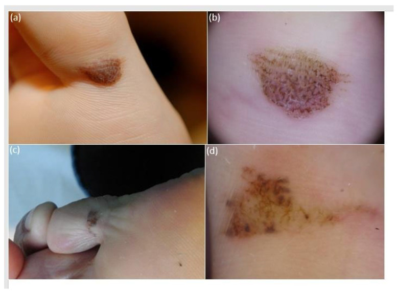
Figure 3.
Clinical appearance of atypical melanocytic plantar lesions of the plantar surface of the fingers, namely first (a) and fifth (c) fingers, presenting as brownish elongated pigmented macules with clear-cut borders and irregular cobblestone-like pigmentation. Lesion one had a maximum diameter of 13 mm and belonged to an 18-year-old female (a). Lesion two had a maximum diameter of 11 mm and belonged to a 52-year-old male (c). Dermoscopic examination (polarized light, 20×) reveals an overall homogenous color arranged both in a parallel furrow and in a cobblestone (b) in case one, which was histologically classified as an acral nevus. Conversely, case two exhibits multiple colors (light brown, dark brown, gray, and reddish) arranged in a multicomponent pattern with streaks, globules, and irregular blotches (d); the lesion was histologically classified as an acral melanoma pt1a.

