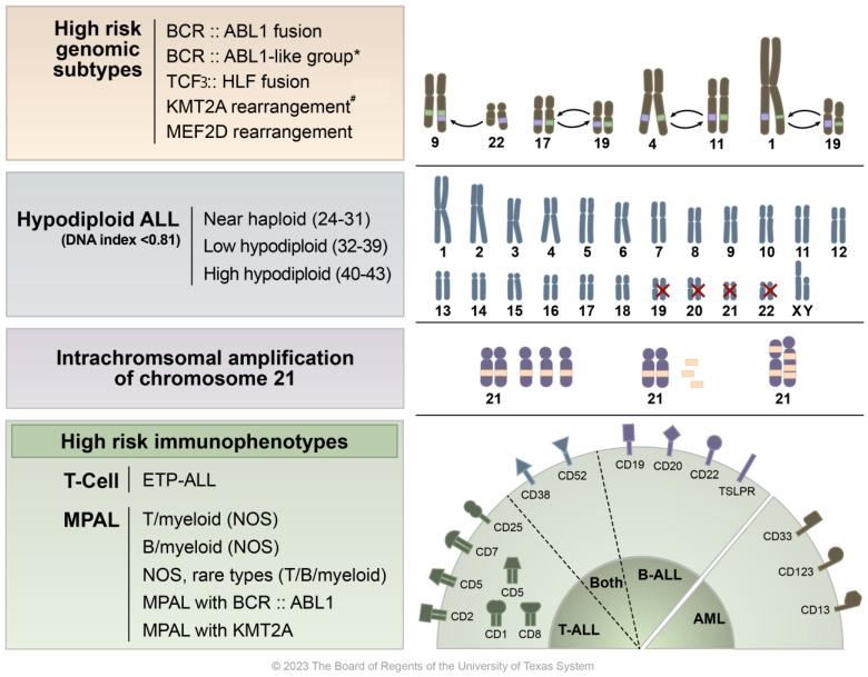
Figure 1.
Overview of high-risk ALL molecular, cytogenetic, and immunophenotypic findings. This figure presents a graphical representation of the various high-risk characteristics found in acute lymphoblastic leukemia (ALL). At the top, high risk genomic features are listed alongside illustrated examples of chromosomal translocations/gene fusions/rearrangements, specifically BCR::ABL1 t(9;22), TCF3::HLF fusion t(17;19), KMT2A rearrangement t(4;11), and MEF2D rearrangement t(1;19). * Although BCR::ABL1-like ALL shares genetic characteristics with Ph+ ALL, it lacks the BCR::ABL1 translocation abnormality. The second row details subtypes of hypodiploid ALL, including one that features the loss of chromosomes 19, 20, 21, and 22. The third row highlights cases where additional copies of a chromosome 21 region, which includes the RUNX1 gene, are present in excess (five or more copies per cell). The bottom row categorizes various high-risk immunophenotypes including early T-cell precursor ALL (ETP-ALL) and mixed phenotype acute leukemia (MPAL), along with their corresponding CD markers. # KMT2A/11q23 has over 100 fusion partners identified, with the most common being t(4;11)—AFF1, t(9;11)—MLLT1, and t(11;19)—MLLT3 (Meyer et al.) [12].

