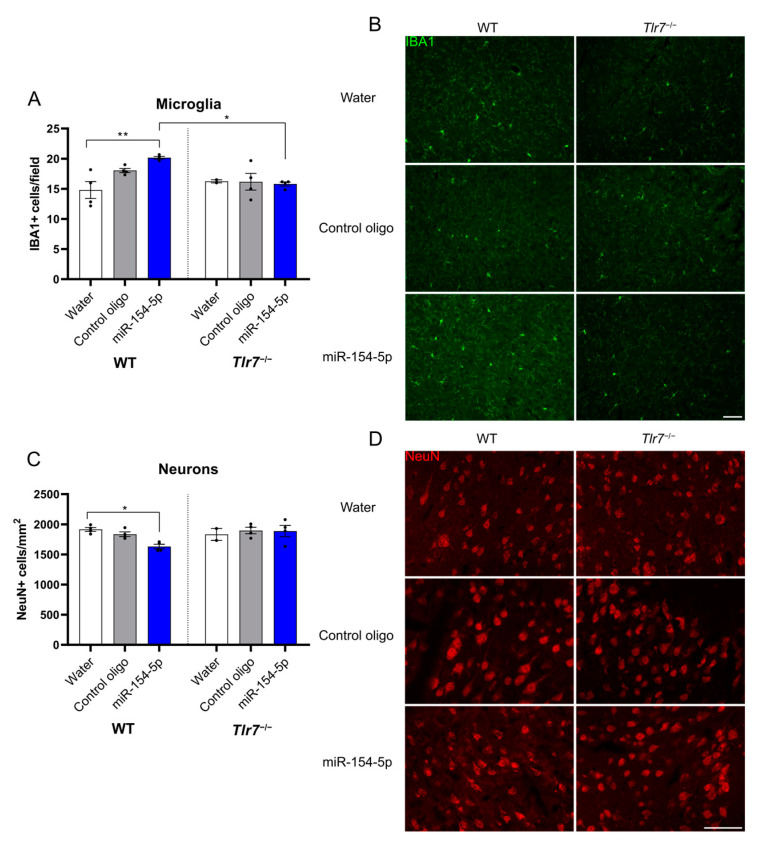
Figure 4.
Intrathecal injection of miR-154-5p into mice induces microglial accumulation and neuronal injury. (A) Mice were intrathecally injected with miR-154-5p (10 μg/mL) or control oligo (10 μg/mL) and sacrificed after 3 days. Sections were immunostained with IBA1 and NeuN antibodies to mark microglia and neurons, respectively. Microglia were quantified in 6 total fields of both hemispheres at 20× magnification. (B) Representative images based on average number of microglia per field. Scale bar represents 50 μm. (C) Average number of NeuN-positive cells in 6 fields of both hemispheres. (D) Representative images based on average number of NeuN-positive cells per field. Scale bar represents 50 μm. Significance was determined by two-way ANOVA followed by Tukey’s multiple comparison test on all conditions to account for both treatment and genetic background. * p < 0.05, ** p < 0.01.

