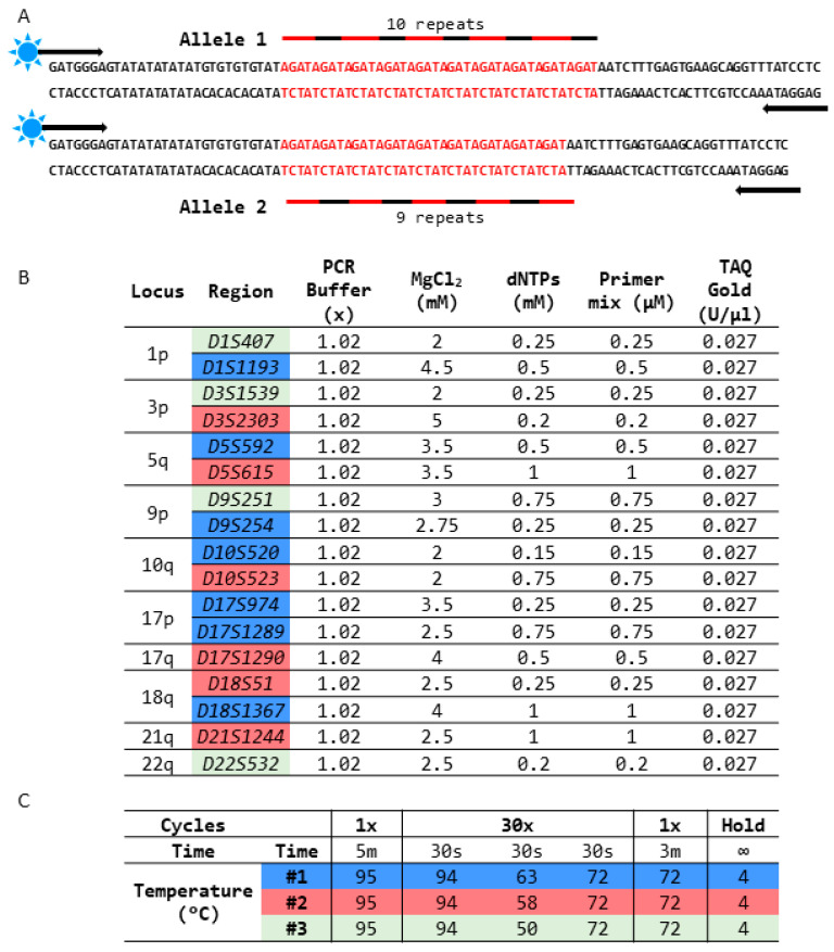
Figure 1.
Design strategy of LOH detection. (A) Representative design shows a sample heterozygous for the D9S254 STR region containing 9 and 10 AGAT repeats. Each STR region is detected by a specific primer pair, where one of the primers is fluorescently tagged and used for detection during capillary electrophoresis. (B) LOH panel markers were analyzed with specific PCR conditions (#1—blue, #2—red, and #3—green) with marker-specific MgCl2 and dNTP concentrations. (C) The 3 PCR conditions used for amplification of the LOH loci are listed and differ in PCR primer annealing temperature. The arrows in the figure indicate direction of primer binding.

