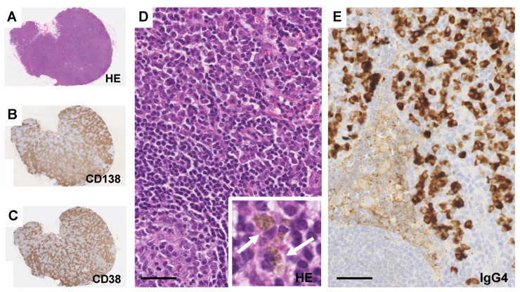
Figure 2.
Light microscopic findings of the lymph node. (A–C) An increase in plasma cells is visible between the enlarged follicles (A), with equal proportions of CD38-positive (B) and CD138-positive (C) plasma cells. (D) A sheet-like plasmacytosis in the interfollicular area. Bar = 50 μm. Insert picture shows scattered hemosiderin-laden macrophages (arrows). (E) Immunostaining of IgG4. Bar = 50 μm.

