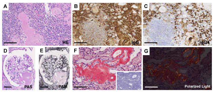
Figure 5.
Light microscopic findings of the kidney. (A) HE staining. Dense plasmacytic infiltration is focally seen in the interstitium. (B,C) Immunostaining of IgG (B) and IgG4 (C). Bars = 100 μm. (D) PAS staining. (E) PAM staining. Nodular deposition of amorphous material is shown in the vascular pole of some glomeruli. Bars = 50 μm. (F) DFS staining. Amorphous deposition in small arteries to arterioles is positive for DFS staining and disappeared after permanganate treatment (insert). Bar = 100 μm. (G) Apple-green birefringence is recognized by polarized microscopy. Bar = 100 μm.

