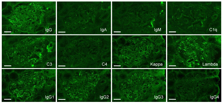
Figure 6.
Immunofluorescence study of the kidney. Relatively faint, granular positivity of IgG and C3 is shown on a glomerular capillary wall. IgA, IgM, C1q, and C4 are negative. IgG subclass staining shows positivity of IgG1, IgG2, and IgG3; however, IgG4 is negative. Light chain restriction is not observed. Bars = 50 μm.

