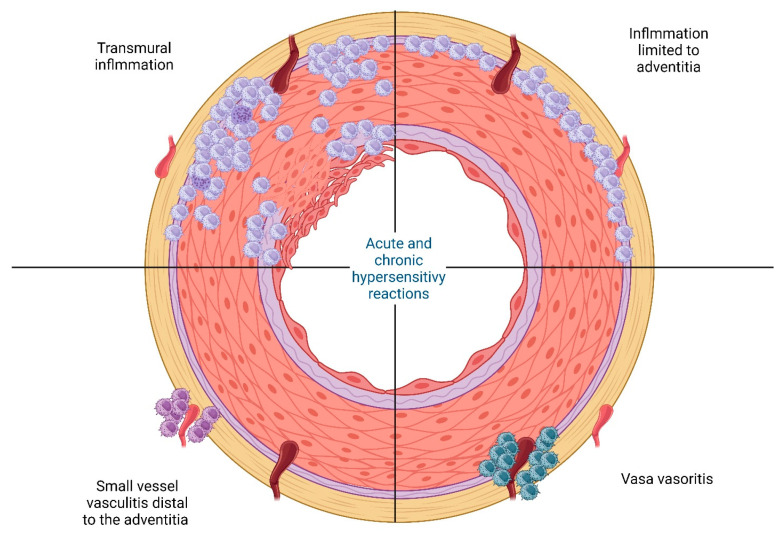
Figure 2.
A visual representation showing the four different histological patterns observed in giant cell arteritis. Upper left: transmural involvement; upper right: Inflammation limited to adventitia; lower left: small vessel vasculitis; and lower right: inflammation limited to vasa vasorum [22]. Created with BioRender.com (accessed on 24 December 2023).

