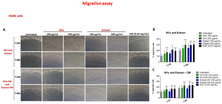
Figure 6.
Effect of NVs and Extract on HUKE cell migration. (A) Migration of HUKE in the agarose spot assay. Spots were left untreated, embedded with EGF, NVs, or Extract at 100 and 200 μg/mL, or conditioned medium (CM) obtained from injured monolayers treated with NVs/Extract, and observed by the microscope 24 h or 48 h later. The dotted white line indicates the spot border. Bar = 100 µm. (B,C) Percentage of migrated cells (distance of the forefront from the border) at different time points was calculated relative to the percentage of migration at t.0 and considered 100%. The results are represented as mean±SD of three experiments. * p < 0.05; *** p < 0.0001 as compared to untreated for each time point.

