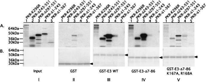
FIG. 7.
Analysis of in vitro interactions between segments of PKR and E3 containing their respective DRBMs. Aliquots of bacterial extracts containing 20 to 100 μg of total protein predetermined to contain similar amounts of GST or GST-E3 fusion proteins (as indicated at bottom of panel B, sections II to V) were combined with an extract prepared from the parental bacterial strain devoid of GST proteins to achieve 200 μg of total bacterial protein. These mixtures were incubated with the 35S-labeled PKR proteins indicated at the top of panel A, section I, and the GST or GST-E3 fusion proteins, along with any bound 35S-labeled PKR proteins, were precipitated by using glutathione-agarose beads and resolved by SDS-PAGE. The 35S-labeled proteins were visualized by autoradiography (A, sections II to V), and the GST or GST-E3 fusion proteins were visualized by Coomassie blue staining (B, sections II to V). Section I in panel A, shows 1/20 (lanes 1 to 4) or 1/10 (lane 5) of the input amounts of 35S-labeled PKR proteins used in the binding assays depicted in sections II to V. Arrowheads in panel B identify GST or the relevant GST-E3 fusion protein.

