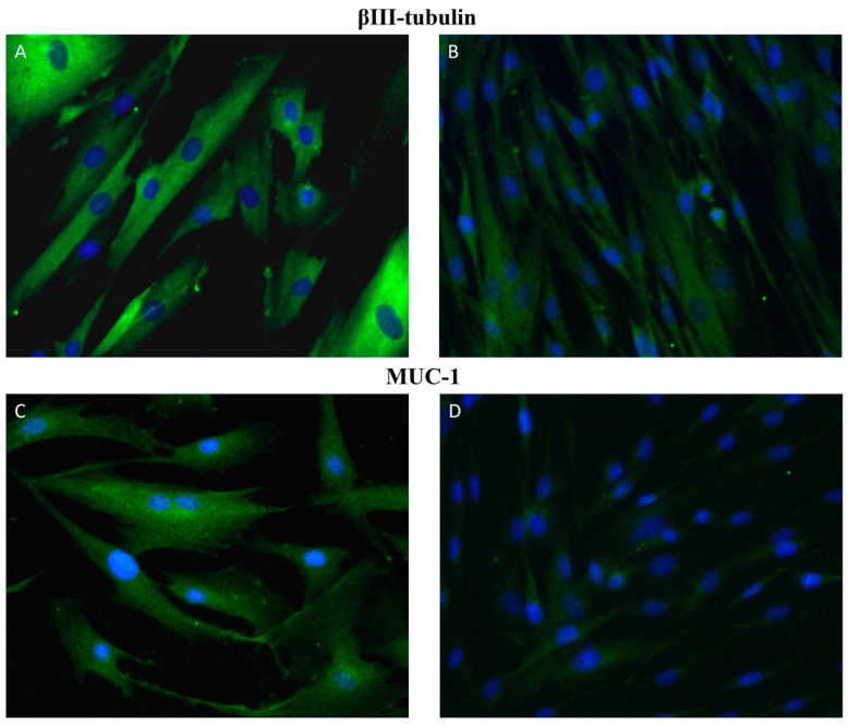
Figure 1.
Immunofluorescence staining of primary cultures of canine mammary glands. Representative images of βIII-tubulin (A,B) and MUC-1 (C,D) positive cells (green). Images (A,C) represent primary cultures isolated from CMT that was histologically diagnosed as tubulopapillary carcinoma of mammary gland. Images (B,C) show healthy non-cancerous cells isolated from canine mammary glands. Magnification: 40×.

