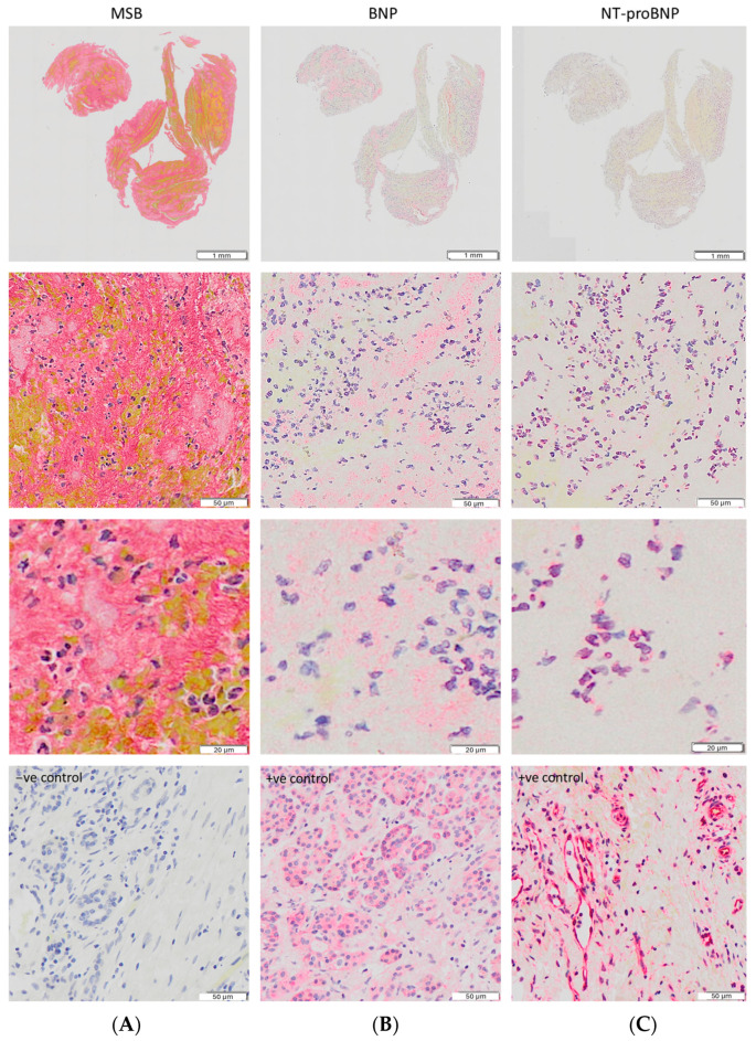
Figure 2.
(A) MSB staining of the main clot components. (B,C) Expression of BNP and NT-proBNP in the same clot. BNP expression is located principally within platelet-rich regions, while NT-proBNP expression is only observed in nucleated cells. Higher magnification images are provided in the third row. In the fourth row, negative staining (A) and positive control staining images (pancreatic cancer tissue) for BNP (B) and proBNP (C) are shown. All images were captured using a 20× objective.

