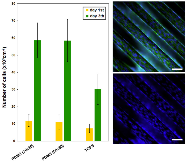
Figure 6.
The number of adhered (day 1) and proliferated (day 3) C2C12 cells cultured on PDMS surfaces (30 × 30 μm, 50 × 50 μm) after collagen coating; tissue polystyrene (TCPS) for comparison is also shown. PDMS surface (line 50 × 50 μm) after collagen coating—fluorescence microscopy images of C2C12 cells with labeled cytoskeletons (in green) and nuclei (in blue) are also shown. White line represents 50 micron.

