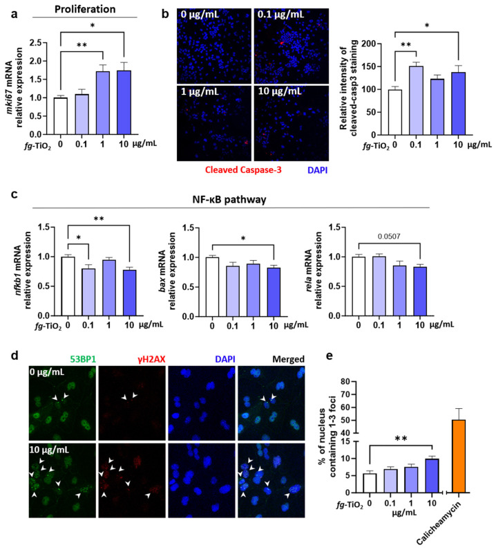
Figure 4.
Effect of fg-TiO2 exposure on cell proliferation, apoptosis, and genotoxicity in EDMs exposed to fg-TiO2 at 0, 0.1, 1, and 10 µg/mL for 24 h. (a) Relative expression of mki67 gene. (b) Immunofluorescence staining of cleaved caspase-3 (20× magnification) and the histogram showing the relative intensity staining compared to control. (c) Relative expressions of genes involved in NF-κB pathway. (d) Immunofluorescence staining of γH2AX and 53BP1 (20× magnification), white arrows pointing foci of γH2AX and 53BP1). (e) Percentage of nucleus containing 1–3 foci of γH2AX and 53BP1. EDMs exposed to calicheamycin γ-1 was used as positive control. Data are presented as the mean ± SEM of three independent experiments, each with four to six EDMs per group. * p < 0.05 and ** p < 0.01 using one-way ANOVA followed by post hoc Dunnett’s multiple comparison test (e) or nonparametric Kruskal–Wallis test, followed by post hoc Dunn’s multiple comparison test (a–c).

