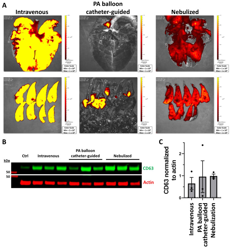
Figure 3.
Pulmonary absorption of exosomes with intravenous, PA balloon catheter-guided, and nebulized delivery. (A) Xenogen imaging of uptake of DiR-labeled PEP in the lungs for intravenous, PA balloon catheter-guided, and nebulized delivery. (B) Western blot demonstrating presence of PEP in control lung (ctrl) tissue compared to lung tissue from intravenous, PA balloon catheter-guided, and nebulized deliveryusing exosomal protein CD63 (green) and loading control actin (red). (C) Quantification (n = 3) of mean fluorescent signal (±SEM) of CD63 normalized to actin loading control; dotted line represents level of control (ctrl).

