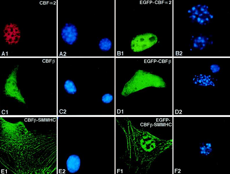
FIG. 2.
Subcellular localization of CBFα2, CBFβ, and CBFβ-SMMHC in transiently transfected cells. NIH 3T3 cells were transfected with pCbfa2 (A1 and A2), pEGFP-Cbfa2 (B1 and B2), pCBFB (C1 and C2), pEGFP-CBFB (D1 and D2), pCBFB-MYH11 (E1 and E2), and pEGFP-CBFB-MYH11 (F1 and F2). Odd-numbered panels show fluorescent signals, while even-numbered panels show DAPI staining of the same fields. Panel A1 shows signal detected with α3043 and a Texas-red-labeled secondary antibody, panels C1 and E1 show signals detected with β141.2 and a fluorescein-labeled secondary antibody, and panels B1, D1, and F1 show the autofluorescent signal of GFP fused to the respective proteins. The antibodies failed to detect the endogenous proteins (compare the untransfected cell in panel A1 with panel A2, as well as that in panel C1 with panel C2).

