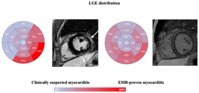
Figure 1.
LGE distribution, according to the American Heart Association bull’s eye plot, in patients with clinically suspected or EMB-proven myocarditis. Focal LGE distribution, mainly located in inferior and lateral walls in clinically suspected myocarditis, as shown by the short axis mid-cavity post-contrast sequence displaying epicardial LGE of the lateral wall (left, white arrow). Diffuse LGE distribution in EMB-proven myocarditis, with evidence of circumferential mid-wall/subepicardial LGE (right).

