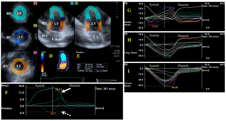
Figure 1.
Evaluation of the left ventricle (LV) by three-dimensional (3D) speckle-tracking echocardiography. Typically, several views are automatically created during the LV assessment using a dedicated vendor-provided software: apical longitudinal four-chamber (A) and two-chamber (B) views and apical (C3), midventricular (C5), and basal (C7) short-axis views. In a 3D LV model (D), LV volumes, ejection fraction, and mass (E) and the apical (white arrow) and basal (white dashed arrow) rotations of the LV (F) are seen with time, as well as the LV global (white curve) and segmental (colored curves) radial (G), longitudinal (H) and circumferential (I) strain curves with time, and an LV volume changes curve with time (dashed white curve) are presented. Abbreviations: RA = right atrium; RV = right ventricle; MASS = LV muscle mass; LA = left atrium; LV = left ventricle; EF = ejection fraction; EDV = end-diastolic volume; and ESV = end-systolic volume.

