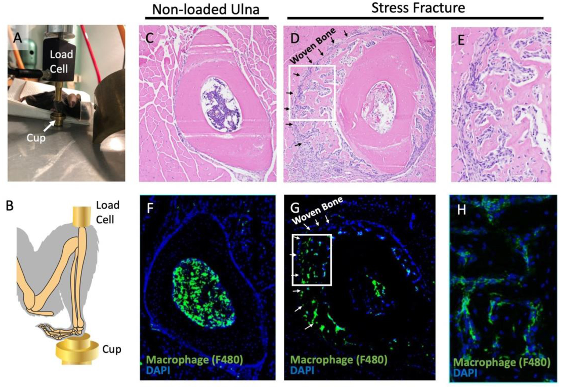
Figure 1. Repeated ulnar loading led to stress fracture and formation of a callus containing macrophages.

(A, B) The mouse was positioned with the ulna in a vertical position between the load cell and cup for stress fracture induction. (C-E) Hematoxylin and eosin staining was performed one week post injury in the ulna when non-loaded (C) or loaded to induce stress fracture (D,E). (F-H) Immunofluorescence with an F4/80 antibody was used to identify macrophages in an unloaded (F) and stress fracture callus (G, H). The boxes in panels D and G outline to the location of panels E and H, respectively. n=2
