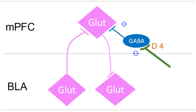
FIGURE 3.

The schematic diagram of the author's estimation of the interrelationships between mPFC neurons and BLA neurons based on the literature of Laviolette et al. 64 ; Wang et al. 65 ; and Floresco and Maric. 66 Glutamate neurons in the mPFC are disinhibited and become more active when inhibitory GABA inter‐neurons are suppressed by the DRD4 effect. mPFC, the medial prefrontal cortex; BLA, basolateral nucleus of the amygdala.
