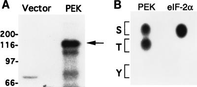
FIG. 4.
PEK in an in vitro kinase assay is autophosphorylated on both serine and threonine residues. (A) Lysates prepared from E. coli expressing PEK or vector alone were incubated with [γ-32P]ATP and analyzed by SDS-PAGE, followed by autoradiography. The radiolabeled PEK is indicated by an arrow on the right. Sizes of protein standards in kilodaltons are on the left. (B) Phosphoamino acid analysis of 32P-labeled PEK from the in vitro assay. Radiolabeled PEK was hydrolyzed with HCl, applied to cellulose-coated sheets, and separated by one-dimensional thin-layer electrophoresis. In parallel, eIF-2α that was phosphorylated by PEK in an in vitro assay was similarly analyzed. The 32P-labeled amino acids were detected by autoradiography. The positions of serine, threonine, and tyrosine are indicated by one-letter codes.

