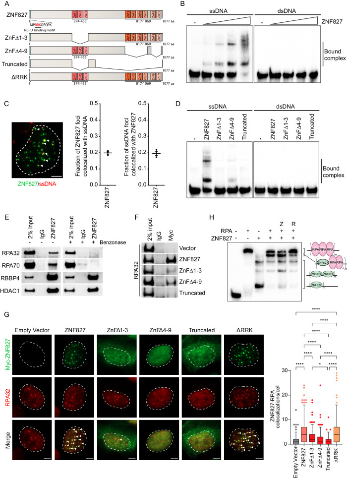
Fig. 1. ZNF827 binds to ssDNA and associates with RPA.
A Schematic of ZNF827 domains and functional mutants. B Electrophoretic mobility shift assay (EMSA) using γ-32P radiolabelled single-stranded and double-stranded pentaprobes with increasing concentrations of purified ZNF827 protein (n = 1). Bound complex is indicated by the gel shift. C Representative image of ZNF827 (green) and native BrdU-stained ssDNA (red) colocalizations in U-2 OS cell nuclei following transient overexpression of ZNF827 (left panel). White arrows indicate colocalizations. Scale bar represents 5 μm. Manual quantitation of ZNF827 and ssDNA foci per nuclei and the fraction of colocalizing foci per nuclei (right panels). Data represent 50 nuclei from three biological replicates. D EMSA using γ-32P radiolabelled single-stranded and double-stranded pentaprobes with purified ZNF827 wild-type and ZNF827 ZnF mutant proteins (n = 1). Bound complex is indicated by the gel shift. E Western blot analysis of RPA components, RPA32 and RPA70, and NuRD components, RBBP4 and HDAC1, following co-immunoprecipitation (co-IP) with a direct ZNF827 antibody in U-2 OS cells overexpressing ZNF827 (n = 1). F Western blot analysis of RPA32 following co-IP with a Myc antibody in U-2 OS cells overexpressing Myc-tagged ZNF827 and ZNF827 ZnF mutants (n = 1). Empty vector included as a negative control. G Representative images of Myc-tagged ZNF827 and ZNF827 mutants (green) and RPA32 (red) immunofluorescence labelling in U-2 OS ZNF827 knockout cells 48 h after transient transfection with Myc-tagged ZNF827 constructs (left panel). White arrows indicate colocalizations. Scale bar represents 5 μm. Manual quantitation of ZNF827 foci colocalizing with RPA foci (right panel). Data represent 50 nuclei from three biological replicates and data from individual nuclei were plotted as Tukey box plots; ****P < 0.0001, *P = 0.05. Multiple comparisons were corrected for using the Bonferroni test. H EMSA using γ-32P radiolabelled single-stranded pentaprobe with purified ZNF827 and RPA protein complex (n = 1). Z denotes addition of ZNF827 15 min before RPA. R denotes addition of RPA 15 min before ZNF827. Bound complex is indicated by the gel shift, and the associated schematic indicates possible protein binding configurations. Source data are provided as a Source Data file.

