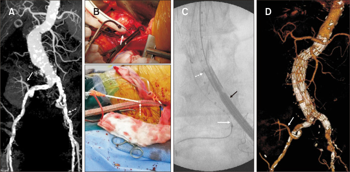
Fig. 3.
Novel strategy for the severely calcified bilateral iliac arteries with transplant kidney. (A) The preoperative maximal intensity projection image showed the abdominal aortic aneurysm and renal artery of a functioning transplanted kidney originating from the right internal iliac artery (arrow). Bilateral iliac arteries showed diffuse, severely calcified stenosis (dotted arrow). (B) (Upper) A 10 mm Dacron graft was anastomosed to the less calcified portion of the iliac artery (arrow). (Lower) Two 7F introducer sheaths (dotted and black arrows) were directly inserted into the graft for the insertion of a sizing pigtail catheter and endograft, respectively. (C) The endograft main body (black arrow) and a sizing pigtail catheter (dotted arrow) were inserted through the temporary conduit. Snaring was successfully used to cannulate the contralateral gate (white arrow). (D) The postoperative volume-rendering image showed no endoleak and a well-perfused transplanted kidney (arrow).

