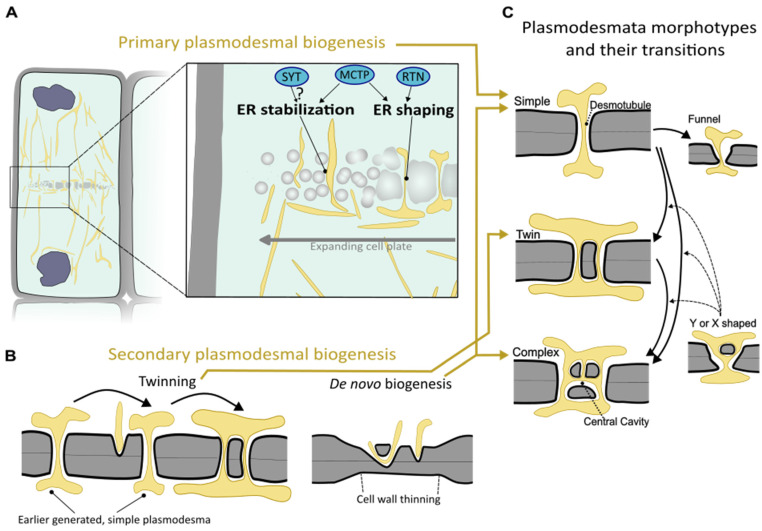
Figure 1.
Schematic depiction of biogenesis of plasmodesmata and plasmodesmata morphotypes. (A) Primary plasmodesmata are synthesized during cytokinesis. A portion of the newly forming cell wall segment termed the cell plate in a dividing cell (left) is shown enlarged on the right. Here, strands of ER traverse the expanding cell plate to ultimately mature into plasmodesmata. In this process, ER strands bridging the expanding cell plate are stabilized by MCTP tethering factors and potentially synaptotagmins (SYTs). ER strands are shaped into the desmotubule, likely by membrane constriction via MCTP and reticulon (RTN)-type proteins. (B) Two means of secondary plasmodesmal biogenesis can be distinguished: a new plasmodesmal channel can be inserted in close proximity to an already existing plasmodesma through a process called ‘twinning’ (left). Furthermore, de novo biogenesis creates plasmodesmata via membrane penetration into an often thinned cell wall segment independent from an existing plasmodesma (right). (C) Different plasmodesma morphotypes and their possible transitions. Simple plasmodesmata can originate from either primary or de novo biogenesis. Simple plasmodesmata can become twinned plasmodesmata via twinning (see (B)). On highly specialized intercellular interfaces, simple plasmodesmata can attain specialized morphologies, such as a funnel-shaped morphology. Complex plasmodesmata are characterized by a central cavity and a branched desmotubule and can arise through the modification of simple or twinned plasmodesmata, but they can also arise during de novo secondary biogenesis. X- or Y-shaped plasmodesmata have an unresolved relationship with the various transitions (being intermediaries during twinning or transformation toward a complex morphotype; dashed arrows). Yellow arrows among (A–C) point to the plasmodesmal morphologies that the biogenic processes can give rise to.

