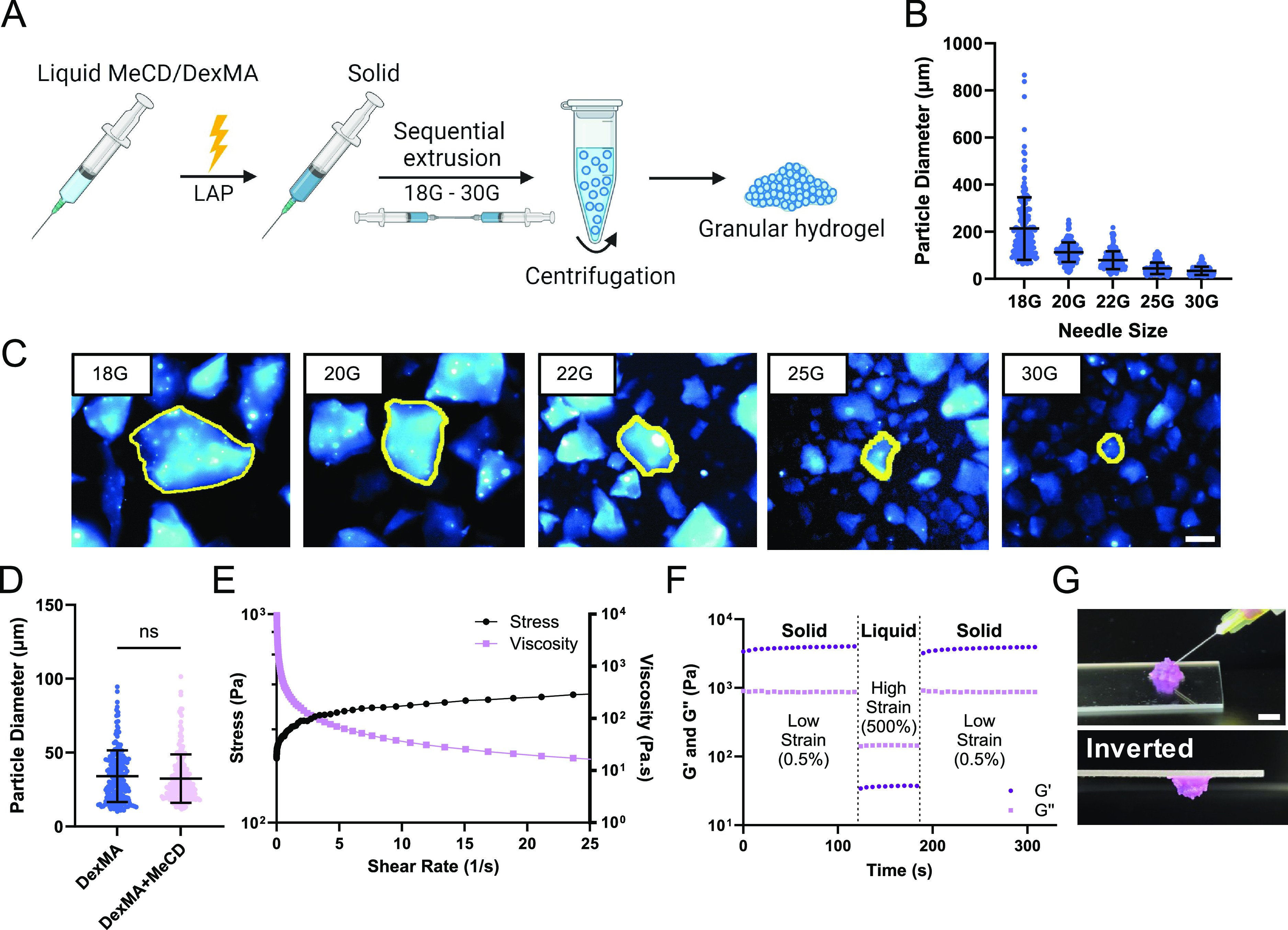
Figure 2.

Granular hydrogel formation and characterization. (A) Schematic representation of microgel fabrication by EF. (B,C) Particle diameter throughout the extrusion process of 5%w/v DexMA gels (mean ± SD; n = 200 particles), quantified using fluorescence microscopy images (C, scale bar = 100 μm). Representative particles are outlined (yellow) for clarity. (D) Final particle diameter of 5%w/v DexMA and 5%w/v DexMA +10%w/v MeCD microgels (mean ± SD; n = 200; ns = not significant). (E) Continuous flow experiments showing the shear stress and viscosity of 5%w/v DexMA +10%w/v MeCD granular hydrogels. (F) Cyclic deformation at low (0.5%) and high (500%) strain (1.0 Hz) of 5%w/v DexMA + 10%w/v MeCD hydrogels; G′ (storage modulus, dark purple, circle), G″ (loss modulus, light purple, circle). (G) Representative images of granular hydrogel injection (30G needle, 1 mL syringe; scale bar = 5 mm).
