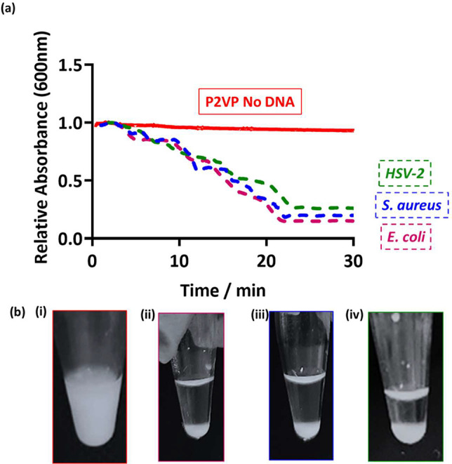
Figure 1.

(a) UV–vis spectrophotometry absorbance at 600 nm as a function of time for PEGMA-P2VP latex only (red) and after the addition of DNA to the latex (HSV-2, green; S. aureus, blue; E. coli, pink). Latex particles were at a concentration of 0.1% w/w and 50 μL of purified amplified PCR product was added. (b) Digital images were taken 30 min after the addition of amplified DNA to 0.2% w/w latex; (i) control with no amplified DNA, (ii) 50 μL of E. coli PCR product, (iii) 50 μL of S. aureus PCR product, and (iv) 20 μL of HSV-2 PCR product.
