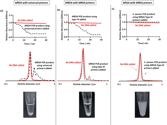
Figure 3.
(Top row) UV–vis spectrophotometry absorbance at 600 nm as a function of time for latex only (red) and after the addition of the PCR product to the latex (black). Latex particles were at a concentration of 0.1 w/w % and 50 μL of purified PCR product was added. (Middle row) DCP particle size distributions obtained for PEGMA-P2VP latex (0.01% w/w) on the addition of PCR products from conventional PCR. (Bottom row) Digital images taken 30 min after the addition of 50 μL of PCR product to 0.2% w/w PEGMA-P2VP latex. For the “MRSA primers” columns, MRSA type III primers were used, giving an overall amplicon size of 280 bp. For the left column, universal bacterial primers were used, yielding an amplicon size of approximately 1400 bp.

