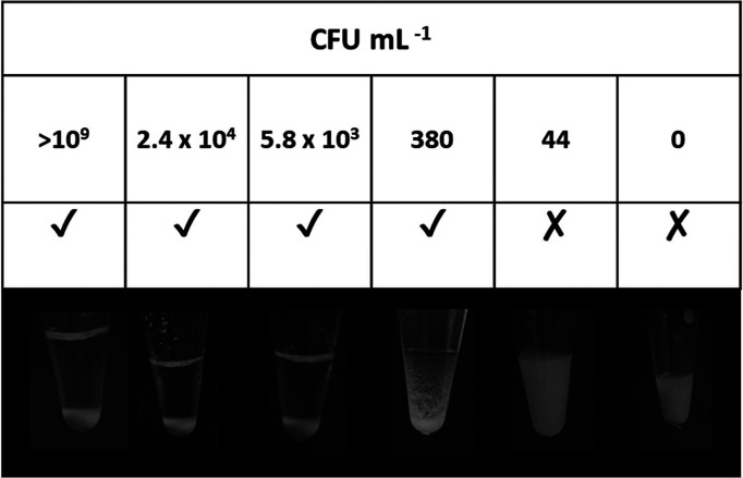
Figure 4.
Digital images showing flocculation of PEGMA-P2VP latexes in the presence of amplified DNA from colony PCR, where the bacterial suspension was diluted by a factor of 10 down to 106 and CFU mL–1 was measured using colony-counting methods prior to amplification via PCR. A tick indicates clear and obvious sedimentation of the latex, a cross indicates that sedimentation did not occur, and a hyphen indicates a borderline result.

