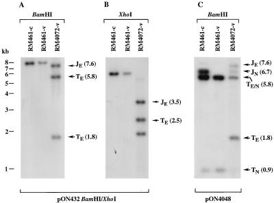
FIG. 3.
Cleavage at the ectopic cleavage site of RM4072. Autoradiograms show cell-associated (-c) and virion (-v) DNAs from the parental virus RM461 and from RM4072 digested with BamHI or XhoI and hybridized with 32P-labeled probes following electrophoresis and transfer to nylon. (A and B) Results of hybridization with a BamHI/XhoI fragment from pON432 which detected fragments from ectopic junctions and termini; (C) results of hybridization with pON4048 DNA to detect fragments from both ectopic and natural cleavage sites and termini. The positions of molecular size markers are shown on the left of panel A, and the locations and sizes in kilobases (in parentheses) of BamHI and XhoI ectopic junction (JE), natural junction (JN), ectopic terminal (TE), natural terminal (TN), and comigrating ectopic and natural terminal (TE/N) fragments are indicated. Note that in panel B, an irrelevant 2.0-kb XhoI fragment adjacent to the ectopic cleavage site also hybridizes to the probe.

