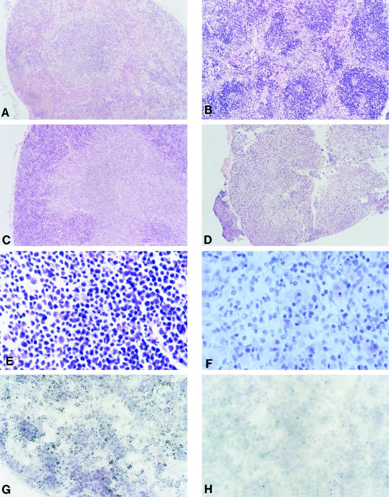
FIG. 2.
Pathology and transgene expression in lymphoid tissues from CD4C/HIVWT Tg mice. (A to F) Light micrographs of various lymphoid tissues. (A and B) Non-Tg and Tg (F17001) spleens, respectively. (C and D) Non-Tg and Tg (F17018) thymuses, respectively. Note the small size of the Tg thymus. (E and F) Non-Tg and Tg (F17001) lymph nodes, respectively. Note the tissue disorganization and hypocellularity of the Tg organ. (G and H) Thymus from a Tg animal (F17001). ISH was performed with antisense (G) or sense (H) probes. Note that in panel G the vast majority of the cells are ISH positive and are of lymphoid morphology. The lymphoid morphology is best shown in panel H on an adjacent section of the same tissue in the absence of a cell-specific hybridization signal when exposed to the sense probe. Magnifications, ×80 (A to D), ×390 (E and F), and ×320 (G and H). The counterstain was hematoxylin and eosin.

